Chinese researchers Dehua Kang, Bin Wang, Yinglin Peng, Xiaowei Liu, and Xiaowu Deng examine methods for improving the bolus, releasing the details of their recent study in ‘Low-Cost iPhone-Assisted Processing to Obtain Radiotherapy Bolus Using Optical Surface Reconstruction and 3D Printing.’
Boluses generally serve as delivery systems, often for medication. In this study, the research is centered around creating a bolus that ‘acts as a water equivalent,’ assisting in providing dose distribution and good coverage. In creating a bolus via an iPhone and desktop 3D printing system, the researchers worked to create more patient-specific systems—fabricated from two CT images of the patient (one for design of the bolus structure; once for dose calculation after the bolus has been created).
Ultimately, a conformal bolus was made with acrylonitrile butadiene styrene (ABS).
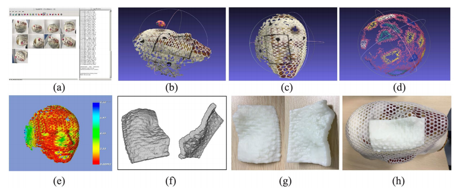
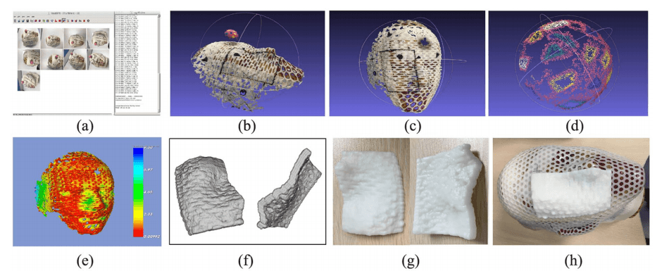
Procedure of bolus reconstruction using the SFM method. (a) The acquired pictures were imported into SFM workspace and 3D reconstruction was run. (b) The surface of the head phantom and the sphere calibration model. (c) The surface of head phantom with a bolus region marker line. (d) The surface of the sphere calibration model. (e) The registration deviation between the two surfaces from the SFM and Marching Cube from CT images. (f) Bolus viewed in the STL format file. (g) The bolus printed using ABS material. (h) The bolus was put in the right place on the head phantom surface.
“The CI and HI of the radiation treatment plans with
“The CI and HI of the radiation treatment plans with patient-specific printed and standard fat bolus were 0.817 and 0.910 (printed) vs 0.697 and 0.887 (fat), respectively,” explained the researchers.
“The prescription dose coverage for PTV in the plan with printed bolus were much better than that in the plan with fat bolus. The V95% (percentage volume received at least 95% of prescription dose) and D95% (dose covered 95% of the volume) in the PTV were 95.65% and 47.96Gy (printed) vs 88.39% and 46.11Gy (fat), while the dose value in every OAR were very similar for the two plans, respectively.”
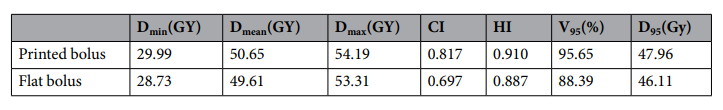
Comparison of HI, CI,V95% and D95% values of PTV between the printed bolus and fat bolus plan.
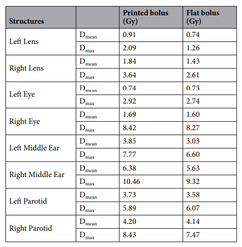
Comparison of parameters of OARs between the printed bolus and fat bolus plan.
“The results demonstrated that the dose coverage and
“The results demonstrated that the dose coverage and conformity of the plan with printed bolus was superior to that with fat bolus, with a higher dose coverage in the superficial PTV area.”

The left panel shows the dose distribution of the 5-beam IMRT plan without a piece of the fat bolus. The right panel shows the dose distribution of the 5-beam IMRT plan with printed bolus. The minimum dose color wash was 4750 cGy. The bottom are the sagittal and coronal views according to the corresponding plans, respectively.
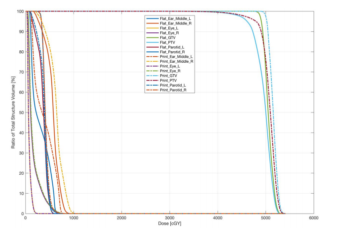
DVH of the two plans with printed bolus and a piece of fat bolus, respectively
In phantom simulation, the researchers noted improved
In phantom simulation, the researchers noted improved distribution of dosage in comparison to the initial bolus, along with better coverage and conformity.
“The V95% for the PTV were 95.65% (3D-printed bolus) vs 88.39% (fat bolus),” said the researchers. “The CI and HI of the plan with 3D-printed bolus raised to 0.817 and 0.910 from 0.697 and 0.887 of that with a fat bolus, respectively.”
Simulation showed that the big air gap decreased as the dose was administered. Image acquisition was easy with the new process, despite the necessity to include the entire target within each picture. Each image also required an overlapping part with the neighboring one—in an effort to decrease the number of scans needed, and patient visits. The researchers noted some of the classic benefits of 3D printing during the process—from accessibility in materials to greater affordability in production.
“For scale the reconstructed structure, a sphere model
“For scale the reconstructed structure, a sphere model with textures of known geometry was used for scale calibration, which ensured accurate 3D reconstruction to design the bolus conformally onto the patient’s irregular body. The radius of the sphere model was set to 15mm because a larger sphere would block the head phantom, while a smaller sphere would lead to low accuracy in the reconstruction result. The 3D sphere fitting algorithm to ft the sphere surface is a robust and accurate method,” concluded the researchers. “The ratio between the fitting radius and the known radius was used as the scaling factor for the reconstructed structure. In the 3D surface scene, it is more difficult and complicated to measure the distance between two points on the irregular surface, but the radius of a sphere can be easily determined using the least square fitting method.”
“The simulation plan shows that the printed bolus was satisfactory for application to improve the dose coverage and conformity in IMRT treatment for a superficial target in the head and neck areas.”
3D printing has been used in a variety of projects involving boluses, from those meant to treat skin cancer, protect cancer patients better during radiotherapy, and further increase effectiveness of treatments. What do you think of this news? Let us know your thoughts! Join the discussion of this and other 3D printing topics at 3DPrintBoard.com.
[Source / Images: ‘Low-Cost iPhone-Assisted Processing to Obtain Radiotherapy Bolus Using Optical Surface Reconstruction and 3D Printing’]
The post Improving Medication Delivery Boluses with an
The post Improving Medication Delivery Boluses with an iPhone & Desktop 3D Printer appeared first on 3DPrint.com | The Voice of 3D Printing / Additive Manufacturing.
from Your daily news from 3DPrint.com https://bit.ly/3dxXyv6