A recent study, conducted by researchers Amber Collings and Katherine Brown at Teeside University in the U.K. analyzed the suitability of specific 3D scanning and modeling techniques in a key aspect of forensic investigation: physical fit analysis (PFA). PFA determines if two pieces of evidence physically fit together, their shared origin, the links between crime scenes and suspects, as well as allowing for object reconstruction for interpretation or presentation, to experts or jurors.
Forensic investigation can be challenging, especially when the evidence is damaged, fragmented, fragile, embedded or hazardous. In PFA, where human bone fragments are manually handled, difficulties arise in matching, reconstructing, interpreting, and presenting the evidence or results of the PFA. The bone fragments may be too small or complex or biologically hazardous. 3D imaging, modeling and printing has already been used broadly to improve or assist in forensics, particularly for complex anthropological evidence. Whether through physical reconstruction or virtual 360 models (built using 3D surface or volume-imaging technique), the use of non-contact 3D technologies has allowed forensics to address issues in visualization, virtual and physical bone reconstruction, matching wounds to weapons, dismemberment and more.
Getting Started with the Tools
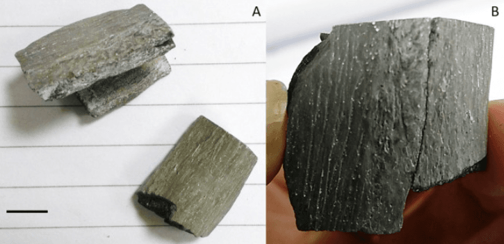
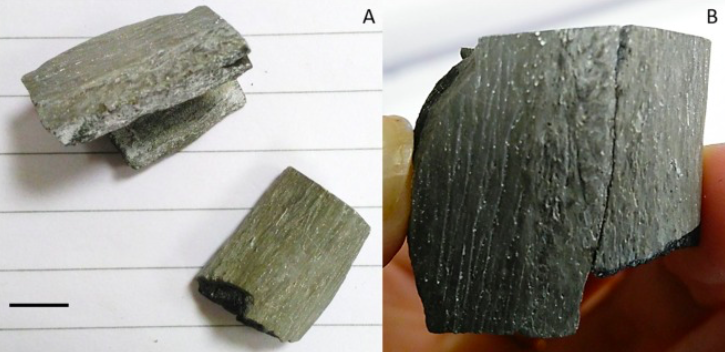
Actual fractured skull bone fragments (A) demonstrating their physical fit (B).
In this study, with burned skull bone fragments as evidence, researchers studied and compared the effectiveness of two imaging techniques to study and 3D print models for use in PFA. Micro computed tomography (µCT) scanning—a more accurate, volume-scanning technique—was compared with structured light scanning, a surface-scanning technique, in terms of effectiveness and efficiency of the virtual model, while fused deposition modeling (FDM) was compared with selective laser sintering (SLS) for the physical 3D model.
While both imaging techniques provided sufficient detail to allow for matching, alignment, and reconstruction, there were differences in efficiency, ease-of-implementation, accuracy and capability. Structure light scanning is cheaper, easier to implement and use, though its lower resolution and limitation in capturing only surface detail meant it was better suited to visualization than fit assessment. It also requires adjustments and sprays to capture the finer details of black or shiny fragments, limiting its ability to be safely handled or used for reconstruction.
µCT provides a far more complete, detailed 3D image, yet requires more expertise and specialized resources. See also: Best 3D Printer Upgrades That Actually Improve Pri…. Since it captures volumetric information it also requires much larger amounts of data. µCT provided better results if a thorough, non-destructive PFA needed to be conducted. Particularly when the evidence involved small, burned bone fragments, in reconstruction of actual or scaled virtual or physical models. Yet structured light scanning proved to be a more feasible alternative for purely visualization purposes.
Step-by-Step Process
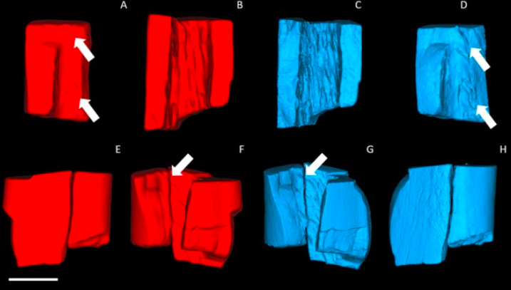
3D models from structured light scanning in red and µCT in blue with fine details.
In terms of 3D printing the models, FDM was a much cheaper alternative to SLS, with the Prusa i3 model (>$1300) priced a hundred times less than laser sintering machines ($5000-175,000). The latter results in far more accurate models and does not require supports but may be prohibitively expensive and requires specialized expertise to operate.
In this study, however, the researchers showed that using FDM was sufficient—by working around issues arising from support structures, hanging features using positioning/orientation, they were able to create accurate, effective 3D models for PFA, thus concluding that, “Fused filament deposition (FFD) 3D printing proved to be an accurate and useful method for creating physical replicas of the bone fragments to perform physical fit analysis (PFA) and bone fragment reconstruction. We therefore recommend μCT imaging paired with FFD 3D printing as an excellent option for non-destructive physical fit confirmation when working with small fragments and burned bone.”
Tips and Tricks for Better Results
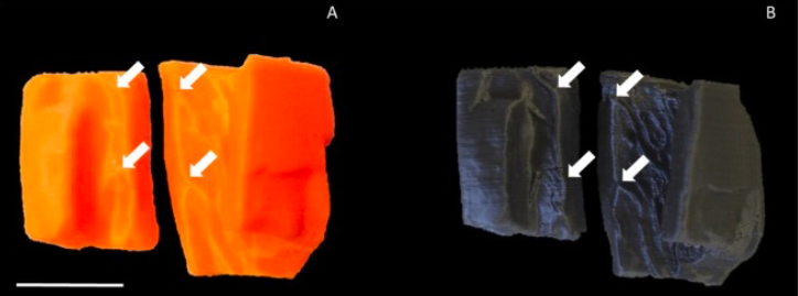
3D printed PLA models – structured light scanned model in orange (A), and µCT in silver (B).
This reaffirms what several studies and recent applications have pushed forward: the combined use of 3D imaging, virtualization and printing can transform or improve forensic investigation, reconstruction, as well as juror understanding in court trials. Some of these applications include developing forensic devices, low-cost postmortem kits, resolving tough to crack cold cases, developing models for forensic training and testing, and more. It is without doubt that implementing 3D technologies significantly improve the speed, efficiency, safety, and capabilities in forensic investigation, thus enabling more accurate, well understood, criminal trial outcomes.
Common Mistakes to Avoid
The post Improving Forensic Analysis of Skull Fragments with 3D Imaging and FDM appeared first on 3DPrint.com | The Voice of 3D Printing / Additive Manufacturing.
from Your daily news from 3DPrint.com https://bit.ly/3hKXgnp
Frequently Asked Questions
What software do I need for 3D modeling?
FreeCAD and Fusion 360 are popular for mechanical parts, Blender for artistic models, and Tinkercad for beginners. Each has different learning curves and capabilities suited to different types of 3D printing projects.
How long does it take to learn 3D modeling for printing?
Basic modeling for 3D printing can be learned in a few days with tools like Tinkercad. More advanced CAD software (FreeCAD, Fusion 360) typically takes 2-4 weeks of regular practice to become proficient for functional parts.
📌 Related Articles
- Best 3D Printer Upgrades That Actually Improve Print Quality: Complete 2026 Guide
- Best Budget 3D Printer Upgrades That Actually Improve Print Quality: Belts, Springs, Hotends & More
- Prusa Research Mini+ vs Prusa MK4: Full Specs Comparison & Buyer’s Guide
- 3D Printing Tungsten Carbide: How Hot-Wire Laser Technology Creates Metal Harder Than Steel
- 3D Printing Safety Equipment Guide: Respirators, Gloves, and Ventilation for 2026