Researchers from China assess the further potential for the smartphone in medical applications, outlining their findings in ‘Evaluating phone camera and cloud service-based 3D imaging and printing of human bones for anatomical education.’
Historically, training on real bodies is one of the best ways for medical students to learn—but cadavers are often not available globally. To make up for that, as technology has progressed, so have a wide range of simulation programs and visual aids. Today, 3D printed medical models and an array of devices are able to offer comprehensive benefits for diagnosing, treating, educating patients, planning for surgeries, and more. Medical students (as well as experienced surgeons) can also benefit enormously from models which may feature tumors about to be operated on in new or rarely performed procedures.
“The primary advantage of 3D printing lies in its ability to create graspable shapes or geometric features of high complexity, overcoming the limitations brought about using flat screens for the visualization of 3D imaging data. Moreover, compared with embalmed cadaveric specimens, 3D printed models are more wear-resistant, easier to clean and store, and, essentially, environmentally green,” state the researchers.
While many industries are benefiting from 3D printing, the
While many industries are benefiting from 3D printing, the impacts are undeniably vast within the medical arena, as anatomical models allow for training and planning, and offer an ‘increasingly significant role’ in developing countries like India.
“In effect, reports indicate that 3D printing helps to improve the effectiveness of teaching and that students learning with 3D printed models performed even better in tests than those learning with real specimens,” stated the researchers.
There are still challenges and limitations in terms of 3D printing as the technology continues to evolve within the medical field; however, education and knowledge of special equipment has been a specific concern. The researchers were motivated in this study to offer technology that is easy to use via phone-based 3D imaging, requiring a basic 3D printer to fabricate models.

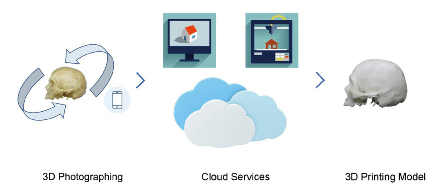
Flow chart of technical
Flow chart of technical route. Three main stages are involved. first, specimens acquired are photographed from all around to obtain enough 2D images from all possible directions. Second, 2D images are converted into digital models with a cloud-based specialized server. Third, after editing, digital models and 3D printing setting data are applied to 3D printer for printing. 2D, two dimensional; 3D, three dimensional.
The following sample bones were used for imaging: femur, rib, cervical vertebra, and skull. Specimens were photographed repeatedly while spinning on a turntable.
“During our testing, the photographer held the phone and captured the images with one hand and rotated the turntable with the other hand after each shot. Two rounds of photography were carried out on different horizontal planes,” explained the researchers.
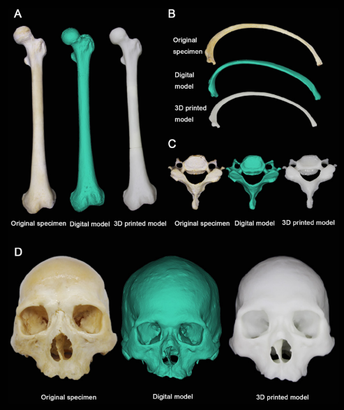
Original specimens, digital models and 3D printed models
Original specimens, digital models and 3D printed models made with SLA technology. (A) femur, (B) rib, (C) cervical vertebra, (D) skull (the digital models may seem smaller because of the special display mode in materialise magics, which is different from single perspective). 3D, threedimensional; SLA, stereolithography apparatus.
Each specimen was photographed 80-100 times, with the photos being uploaded to Get3D and the converted files sent to an online 3D printing service in China. The researchers stated that the costs of the 3D printed femur, rib, cervical vertebra, and skull were USD $20.27, $3.96, $1.13, and $35.40, respectively.
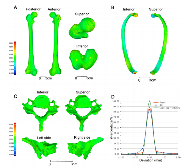
Analysis of deviation between original specimens and 3D printed models. Gradation on the deviation spectrum is 0.5mm each; green colour indicates deviations ranging from −0.5 to 0.5mm; hot colour indicates positive deviations ranging from 0.5 to 2mm; cool colour indicates negative deviations ranging from −0.5 to 2mm. deviation analysis of (A) femur, (B) rib, (C) cervical vertebra; (D) distribution of deviations (in %). 3D, three-dimensional.

Analysis of the deviation between digital models and 3D printed
Analysis of the deviation between digital models and 3D printed models. Gradation on the deviation spectrum is 0.5mm each; green color indicates deviations ranging from −0.5 to 0.5mm; hot color indicates positive deviations ranging from 0.5 to 2mm; cool color indicates negative deviations ranging from −0.5 to 2mm. deviation analysis of (A) femur, (B) rib, (C) cervical vertebra; (D) skull; (E) distribution of deviations (in %). 3D, three-dimensional.
Upon evaluation, the researchers confirmed that their method offered a ‘fairly high precision’ in digital and 3D printed models.
“The most noteworthy feature of the proposed workflow is that it works without scanners or the CT/MRI dataset, thus enabling a broader range of 3D printing technology for educational applications,” stated the authors.
The models offered a good display of anatomical features;
The models offered a good display of anatomical features; for example, the nutrient foramina on the femur was ‘observed easily.’ This was noted in comparison to previous research where an FDM 3D printer using ABS rendered a similar femur sample to be invisible in the area of the nutrient foramina. For this study, SLA was chosen, with Somos Imagine 8000—a rigid material that is tough, dense, and easy to clean.
“The 3D printed models created using the photogrammetry method demonstrate only the external features of the bone specimens; the inner structures are invisible. Human specimens also have this limitation. To display the different anatomical landmarks on the interior of the skull, three or four differently dissected specimens must be used. The same strategy can be applied while creating 3D printed models that display different anatomical structures—differently dissected or sliced specimens are chosen as the resources to be put through photogrammetry,” explained the researchers in their final discussion.
“The photogrammetric digitization workflow adapted in the present study demonstrates fairly high precision with relatively low cost and fewer equipment requirements. This workflow is expected to be used in morphological/anatomical science education, particularly in institutions and schools with limited funds or in certain field research projects involving the fast acquisition of 3D digital data on human/animal bone specimens or on other remains.”

Comparison of fine structures among specimens, digital models and
Comparison of fine structures among specimens, digital models and prints. (A) Nutrient foramina in the great trochanter (above) and the fovea for ligament of head (below). (B) Nerve foramina in cranial base (above) and intraorbital structures (below). (C) Tubercle of the rib. (D) Nutrient foramina in vertebral body
What do you think of this news? Let us know your thoughts! Join the discussion of this and other 3D printing topics at 3DPrintBoard.com.
[Source / Images: ‘Evaluating phone camera and cloud service-based 3D imaging and printing of human bones for anatomical education’]
The post China: Smart Phones as Imaging Devices for Human Bones to be 3D Printed & Used in Education appeared first on 3DPrint.com | The Voice of 3D Printing / Additive Manufacturing.
from Your daily news from 3DPrint.com http://bit.ly/3cHLwj4