Quick Answer: What is Cryogenic 3D Printing for Bone Regeneration?
Cryogenic 3D printing is an advanced bioprinting technique that uses low temperatures to create scaffolds for bone regeneration. Unlike traditional bioprinting methods, cryogenic printing freezes the material as it’s deposited, allowing for the creation of extremely soft yet stable structures that can support cell growth. This technique is particularly effective for bone regeneration because it can create scaffolds that closely match the mechanical properties of human bone while delivering growth-promoting peptides in a controlled manner.
Introduction: The Future of Bone Regeneration
Researchers from China have made significant strides in bone regeneration technology with their groundbreaking study on cryogenic 3D printing. Published in “Cryogenic 3D printing of dual-delivery scaffolds for improved bone regeneration with enhanced vascularization,” this research addresses one of the most persistent challenges in tissue engineering: creating structures that can both stimulate bone growth and support blood vessel formation simultaneously.
Bone regeneration has been a priority area in biomedical research for decades, particularly as populations age and the demand for effective treatments increases. The field has evolved dramatically in the last five years, with 3D printing becoming increasingly mainstream and bioprinting techniques maturing rapidly. Despite this progress, achieving both osteogenesis (bone formation) and vascularization (blood vessel formation) in regenerated tissue remains a significant hurdle.
Understanding the Challenge of Bone Regeneration
Successful bone regeneration requires more than just filling a gap in bone tissue. It demands a sophisticated approach that considers multiple biological processes working in harmony:
- Cell Viability: The biggest challenge in tissue engineering is keeping cells alive not just during growth but throughout the entire regeneration process. Cells are delicate and require specific conditions to thrive.
- Mechanical Compatibility: Regenerated bone must match the mechanical properties of surrounding natural bone to prevent stress shielding or failure under load.
- Vascular Integration: New bone tissue needs a blood supply to deliver nutrients and oxygen. Without proper vascularization, regenerated tissue cannot survive long-term.
- Structural Support: Scaffolds must provide temporary support while new tissue forms, then degrade safely without causing inflammation or other complications.
Traditional bone regeneration methods often struggle to balance these competing requirements. Autografts (using a patient’s own bone) are limited by donor site availability and morbidity, while allografts (using donor bone) carry risks of disease transmission and immune rejection. Synthetic materials offer control but often lack biological functionality.
The Innovation: Dual-Peptide Delivery via Cryogenic Printing
The Chinese research team’s breakthrough lies in their use of cryogenic 3D printing to create scaffolds that deliver two different peptides with complementary functions:
- Osteogenic Peptide (OP): Promotes osteogenesis—the formation of new bone tissue. This peptide stimulates osteoblasts (bone-forming cells) to produce new bone matrix.
- Angiogenic Peptide (AP): Promotes angiogenesis—the formation of new blood vessels. This peptide encourages endothelial cells to form vascular networks that will supply blood to the regenerated bone.
By combining these two peptides in a single scaffold, the researchers created a synergistic system where each peptide supports the other’s function. Blood vessels deliver nutrients needed for bone growth, while new bone provides structure for vascular networks to develop.
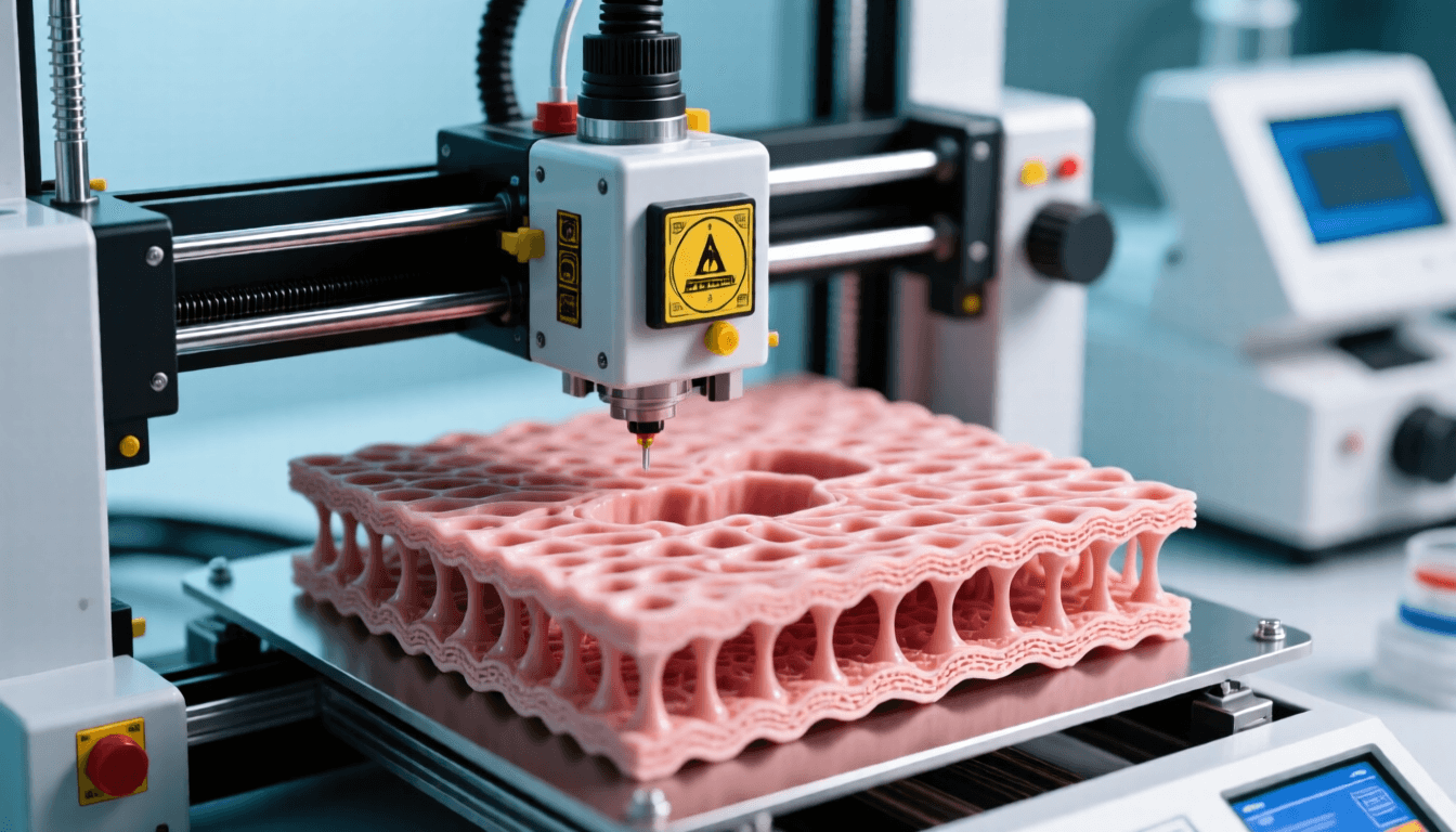
How Cryogenic 3D Printing Works
Cryogenic 3D printing differs from conventional bioprinting in several key ways. The process involves several critical steps:
Step 1: Material Preparation
The researchers used β-tricalcium phosphate (β-TCP), a calcium phosphate ceramic widely used in bone regeneration due to its biocompatibility and similarity to natural bone mineral. This material was agitated for 30 minutes in an ice water bath—a crucial step that gives cryogenic printing its name. The cold temperature initiates phase changes that will later enable precise structure formation.
Step 2: Peptide Integration
The osteogenic peptide was mixed with the TCP material using composite emulsion inks. These specialized inks allow peptides to be evenly distributed throughout the scaffold material and released in a controlled manner over time.
Step 3: Printing Process
Using a specialty 3D printer, the peptide-loaded TCP was deposited in precise patterns. The cryogenic environment ensured that the material maintained its shape as it was printed, overcoming one of the main challenges in printing soft, biologically active materials—preventing collapse or deformation during the fabrication process.
Step 4: Freeze Drying
After printing, scaffolds were freeze-dried to remove water and composite emulsion inks. This process preserved the delicate structure of the scaffold while removing materials that could interfere with later peptide coatings.
Step 5: Angiogenic Peptide Coating
Scaffolds were then coated with angiogenic peptide contained within collagen. This coating was gelled for 30 minutes at 37°C (body temperature) and then dried at room temperature. The collagen carrier ensures that AP is released in a controlled manner that complements the OP release from the scaffold core.
Key Results: Improved Regeneration and Vascularization
The dual-peptide delivery approach produced remarkable results in both laboratory and animal testing:
In Vitro Performance
Laboratory tests showed distinct release profiles for the two peptides. The angiogenic peptide was released rapidly, reaching 58% within the first few days before plateauing. This immediate release helps establish vascular networks early in the regeneration process. In contrast, the osteogenic peptide showed more sustained release, reaching 79% after 42 days, providing long-term support for bone formation.
In Vivo Success
Animal studies using rat cranial defects demonstrated the effectiveness of the approach. Scaffolds containing only angiogenic peptide (TV scaffolds) showed incomplete regeneration, while those containing only osteogenic peptide (TB scaffolds) produced better bone formation. However, the dual-peptide scaffolds (TVB) showed the greatest improvement, with complete regeneration of cranial defects.
Mechanical Properties
Mechanical testing revealed that the scaffolds closely matched the properties of human bone. The collagen coating reduced compressive strength slightly, but this was deemed acceptable for bone tissue engineering applications. The scaffolds maintained enough structural integrity to support cell growth while still being suitable for eventual degradation and replacement by natural bone.
Comparison: Cryogenic vs. Conventional Bioprinting Methods
| Feature | Cryogenic 3D Printing | Conventional Bioprinting |
|---|---|---|
| Temperature During Printing | Low temperature (often below freezing) | Room temperature or body temperature |
| Material Stability | High—structures freeze in place immediately | Moderate—materials may flow or deform |
| Complexity of Structures | Can create very soft, delicate structures | Limited by material viscosity and stability |
| Cell Viability | Excellent—reduced thermal stress | Good, but sensitive to temperature changes |
| Peptide Loading | High—stable structures can carry more biological cargo | Moderate—limited by structural stability |
| Cost | Higher—requires temperature control | Lower—standard equipment sufficient |
Comparison: Different Scaffold Materials for Bone Regeneration
| Material | Advantages | Disadvantages | Best Applications |
|---|---|---|---|
| β-Tricalcium Phosphate (β-TCP) | Excellent biocompatibility, similar to bone mineral, supports osteogenesis | Can be brittle, slower degradation rate | Load-bearing bone defects, large defects |
| Hydroxyapatite | Very similar to natural bone mineral, excellent osteoconductivity | Very slow degradation, brittle | Dental applications, coating implants |
| Collagen | Biologically active, supports cell attachment, biodegradable | Low mechanical strength, rapid degradation | Soft tissue applications, coating materials |
| Polycaprolactone (PCL) | Good mechanical strength, slow degradation, easy to print | Hydrophobic, lacks biological activity | Structural scaffolds, long-term implants |
| Dual-Peptide Scaffolds (Study) | Combines osteogenic and angiogenic effects, controlled release | Complex manufacturing, higher cost | Complex bone defects requiring vascularization |
Future Implications and Applications
The success of this study opens numerous possibilities for clinical applications and future research:
Clinical Translation
While the research was conducted on rat models, the principles demonstrated are applicable to human bone regeneration. Potential clinical applications include:
- Dental Implants: Improving integration of dental implants with surrounding bone
- Spinal Fusion: Enhancing bone growth between vertebrae
- Fracture Repair: Accelerating healing of complex fractures
- Joint Reconstruction: Supporting bone growth in joint replacement procedures
Customization and Personalization
3D printing enables patient-specific scaffolds that match the exact geometry of bone defects. This personalized approach could improve outcomes and reduce complications. The cryogenic method’s ability to create complex, soft structures without collapse makes it particularly suitable for irregular defect shapes.
Expanded Therapeutic Applications
The dual-delivery concept could be applied beyond bone regeneration:
- Cartilage Repair: Using different peptides to promote cartilage formation and lubrication
- Tendon/Ligament Regeneration: Delivering peptides that support connective tissue growth
- Nerve Regeneration: Combining neurotrophic factors with structural support
Challenges and Considerations
Despite the promising results, several challenges remain before widespread clinical adoption:
Regulatory Approval
Medical devices involving biological materials face rigorous regulatory scrutiny. The combination of multiple active ingredients (OP, AP) in a single scaffold complicates the approval process. Each component must be demonstrated safe and effective independently and in combination.
Manufacturing Scale-Up
Transitioning from laboratory-scale production to commercial manufacturing requires addressing quality control, consistency, and cost-effectiveness. Cryogenic printing’s need for precise temperature control adds complexity and cost to production.
Long-Term Outcomes
While animal studies showed excellent results over three months, human applications may require longer-term monitoring to ensure continued success and absence of delayed complications.
Related Research in Bone Regeneration
This study is part of a broader wave of innovation in bone regeneration research. Other notable approaches include:
- Titanium Additive Manufacturing: Using 3D-printed titanium scaffolds for large bone defects. See also: Best Budget 3D Printer Upgrades That Actually Impr…. Titanium offers excellent strength and biocompatibility but lacks biological activity.
- Halloysite Nanotubes: Louisiana Tech researchers have explored incorporating halloysite nanotubes into bone regeneration scaffolds to improve mechanical properties and enable controlled drug delivery.
- Hydroxyapatite Structures: Various groups are investigating 3D-printed hydroxyapatite structures for bone repair, capitalizing on hydroxyapatite’s similarity to natural bone mineral.
Conclusion: A Step Toward Better Bone Healing
The Chinese researchers’ work on cryogenic 3D printing with dual-peptide delivery represents a significant advance in bone regeneration technology. By combining precise manufacturing techniques with biological insight, they’ve created scaffolds that address two critical aspects of regeneration: bone formation and blood vessel growth.
While clinical translation will take time and require further study, the approach demonstrates the power of combining engineering principles with biological understanding. As 3D printing technology continues to advance and our understanding of regenerative medicine grows, techniques like cryogenic dual-peptide delivery may become standard tools in the orthopedic surgeon’s repertoire.
For patients facing bone loss from trauma, disease, or congenital conditions, these advances offer hope for more effective, less invasive treatments. The ability to regenerate bone using scaffolds that closely mimic natural bone’s composition and function could transform outcomes in a wide range of clinical scenarios.
Frequently Asked Questions
Q1: What is cryogenic 3D printing and how does it differ from regular 3D printing?
A: Cryogenic 3D printing uses low temperatures (often below freezing) during the printing process. Unlike regular 3D printing, which typically operates at room or elevated temperatures, cryogenic printing causes materials to freeze or solidify as they’re deposited. This allows for the creation of very soft, delicate structures that would collapse under normal printing conditions. The cold temperature also helps preserve biological activity in sensitive materials like peptides and cells.
Q2: Why is vascularization so important for bone regeneration?
A: Vascularization (the formation of blood vessels) is critical because regenerated bone tissue needs a blood supply to receive oxygen, nutrients, and growth factors while removing waste products. Without proper vascularization, new bone cells cannot survive long-term, and the regenerated tissue will fail. Blood vessels also provide pathways for immune cells and stem cells to reach the regeneration site, supporting the healing process.
Q3: How long does it take for cryogenic 3D-printed scaffolds to regenerate bone?
A: The study showed significant bone regeneration within three months in rat models. However, the timeline can vary depending on several factors: the size and location of the bone defect, the patient’s age and overall health, the specific scaffold design used, and whether additional growth factors or cells are included. In clinical applications, complete regeneration may take 6-12 months or longer, though initial integration can occur within weeks.
Q4: Are there any risks associated with using peptide-loaded scaffolds for bone regeneration?
A: Like any medical intervention, there are potential risks to consider. These include possible immune reactions to the peptides or scaffold materials, infection at the implantation site, and potential for improper degradation leading to inflammation. The study showed promising results with no adverse effects in animal models, but human trials would be needed to fully assess safety. Additionally, the controlled release of peptides must be carefully calibrated—too much or too little could affect outcomes.
Q5: Can cryogenic 3D printing be used for applications other than bone regeneration?
A: Yes, the principles demonstrated in this bone regeneration study have applications beyond orthopedics. The ability to create soft, stable structures that deliver multiple biological factors in a controlled manner makes cryogenic printing suitable for other tissue engineering applications, including cartilage repair, tendon and ligament regeneration, and potentially even organ printing. The dual-delivery concept could be adapted for various tissue types by selecting appropriate growth factors, peptides, or other biological agents.
Q6: How does this technology compare to traditional bone grafts?
A: Cryogenic 3D-printed scaffolds offer several advantages over traditional bone grafts. Autografts (using a patient’s own bone) are limited by donor site availability and can cause significant pain and complications at the harvest site. Allografts (donor bone) carry risks of disease transmission and immune rejection. Cryogenic-printed scaffolds eliminate the need for donor tissue, can be customized to fit any defect shape, and can be engineered to release growth factors in a controlled manner—something natural bone grafts cannot do. However, natural bone grafts have the advantage of being fully biologically active from the start, whereas scaffolds may require time to integrate.
Q7: When might this technology be available for clinical use in humans?
A: While the results are promising, clinical translation typically takes several years. The technology would need to undergo extensive testing in larger animal models, followed by human clinical trials to demonstrate safety and efficacy. Regulatory approval from agencies like the FDA (in the US) or EMA (in Europe) would also be required. Given the complexity of the dual-peptide delivery system, it’s realistic to expect clinical trials to begin within 3-5 years, with potential market availability in 7-10 years if trials are successful. However, timelines can vary significantly based on research findings and regulatory pathways.
Sources:
Related: Improved Bioprinting for Jaw Bone Regeneration with Controlled Release, Antibact · 3D Printing: Successful Scaffolds in Bone Regeneration · Tissue Engineering for Bone Regeneration: 3D Printing of Piezoelectric Barium Ti
- Cryogenic 3D printing of dual-delivery scaffolds for improved bone regeneration with enhanced vascularization. ScienceDirect.
- Bone regeneration techniques and 3D printing applications. 3DPrint.com.
- Louisiana Tech: 3D printing halloysite nanotubes for improved bone regeneration methods.
- Researchers testing 3D printed hydroxyapatite structures for bone regeneration. 3DPrint.com.
- Cryogenic 3D printing at low temperatures for tissue engineering. Mary Ann Liebert, Inc.
- Freezing printed semi-liquid structures in cryogenic fabrication. MDPI Materials.
Frequently Asked Questions
How is 3D printing used in medicine?
3D printing is used in medicine for surgical planning models, custom implants, bioprinting tissue scaffolds, drug delivery systems, dental aligners, and prosthetics. It enables patient-specific solutions that improve outcomes and reduce surgery time.
What materials are biocompatible for 3D printing?
Common biocompatible materials include PEEK, titanium alloys (Ti6Al4V), bio-ceramics (hydroxyapatite), medical-grade resins, PLA for temporary implants, and hydrogels for bioprinting. Material choice depends on the application and required mechanical properties.
Is 3D printed medical equipment FDA approved?
Yes, several 3D printed medical devices have FDA clearance, including orthopedic implants, dental restorations, and surgical guides. Each device must go through the appropriate regulatory pathway based on its risk classification.
📌 Related Articles
- Best 3D Printer Upgrades That Actually Improve Print Quality: Complete 2026 Guide
- Best Budget 3D Printer Upgrades That Actually Improve Print Quality: Belts, Springs, Hotends & More
- 3D Printing Safety Equipment Guide: Respirators, Gloves, and Ventilation for 2026
- Prusa Research Mini+ vs Prusa MK4: Full Specs Comparison & Buyer’s Guide
- Bambu Lab P1S vs Bambu Lab P2S: Full Specs Comparison & Buyer’s Guide