Researchers from China are exploring the feasibility of 3D printing for pre-planning of complex surgeries, detailing the results of their study, involving the data of 124 patients, in the recently published ‘Three-dimensional printing in the preoperative planning of thoracoscopic pulmonary segmentectomy.’
The overall goal of the study was to compare 3D printing in pre-operative care to three-dimensional computed tomography (3D-CT) in thoracoscopic pulmonary segmentectomy. These procedures are meant to treat early-stage lung cancer and are expected to be used even more in the future to eradicate lung cancer caught in the beginning stages.
The lung segment is much more complex than the pulmonary lobe, leaving surgeons to seek out better techniques for seeing the anatomy involved as well as guiding the surgery. The lobectomy is still the traditional method of choice, however, and using the pulmonary segmentectomy as treatment is considered controversial by some. Such a procedure also requires greater skill in the operating room, as well as more experience in navigating surgery of the lung segment. Without comprehensively distinguishing the structure of the pulmonary segment before operating, the authors state that injuries are likely to be caused.
“Three-dimensional computed tomography (3D-CT) can well reconstruct the 3D images of vessels, bronchi, and tumor of the patient’s diseased pulmonary lobe and now is widely used to make a preoperative plan,” state the researchers. “However, 3D-CT still shows 3D images on 2D screens, which still has great limitations in viewing the anatomy of pulmonary vessels and bronchi.”
Understanding Filament Properties
Patients were divided into three groups for the study: General, 3D-CT, and 3D printing. Pre-operative 3D image reconstruction was performed to view and reconstruct 3D images of the nodules, bronchi, and pulmonary vessels. The models were then printed on a Lite600HD 3D printer.

Material Comparison and Selection
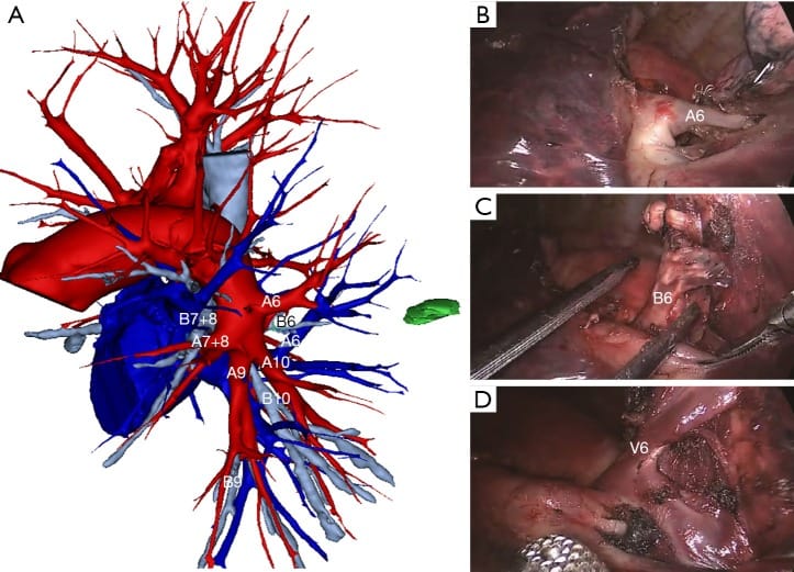
Photos from thoracoscopic LS6 segmentectomy. (A) Reconstructing 3D-CT image for LS6 segmentectomy; (B) pulmonary artery of LS6; (C) bronchus of LS6; (D) pulmonary vein of LS6.
As the actual surgery progressed, the patients received general anesthesia. Two to three incisions were made during the procedure, with camera ports in place. On point with the 3D-CT image or 3D printing model, the authors noted that ‘the arteries and veins of the target segment were found carefully during the operation.’

Print Settings and Optimization
Photos from thoracoscopic RS9 segmentectomy. (A) 3D printed model of vessels and bronchi of the right lobe; (B) 3D printed model of arteries, veins, and bronchi of the right low lobe; (C) pulmonary arteries of RS9; (D) pulmonary arteries of RS9; (E) pulmonary vein of RS9; (F) bronchus of RS9. RPA, right pulmonary artery; RPB, right pulmonary bronchus; RPV, right pulmonary vein.
Highlights from the study showed the following:
- Intraoperative blood loss in 3D-CT group and 3D printing group decreased significantly
- Operation time in 3D-CT group and 3D printing group was significantly shorter than in the general group
- Differences in operation time for general group were significantly longer
Strength and Durability Testing
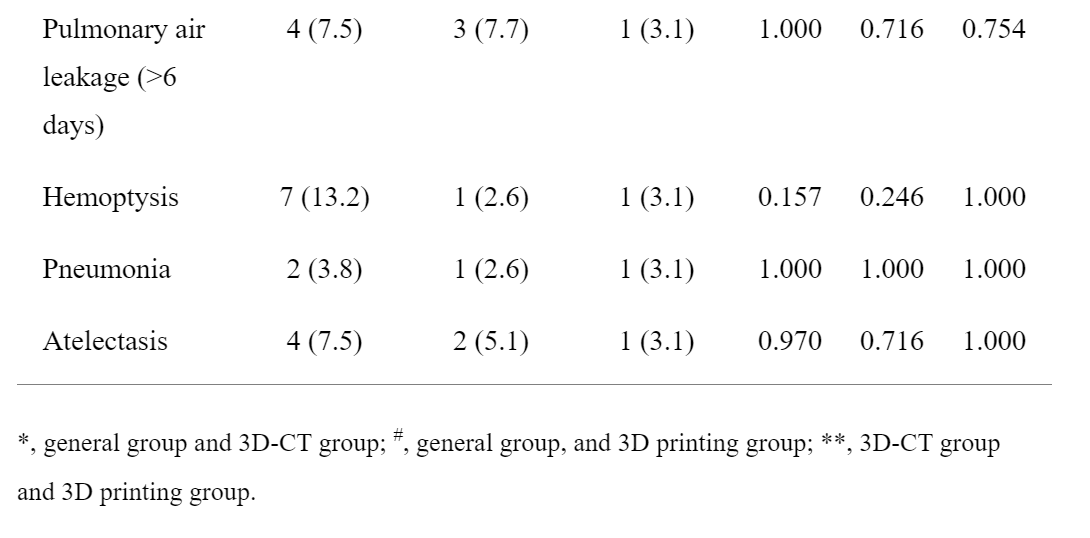
*, general group and 3D-CT group; #, general group, and 3D printing group; **, 3D-CT group and 3D printing group.
One patient (from the 3D-CT group) experienced intraoperative injury, requiring thoracotomy; however, the researchers point out there was no conversion necessary in the 3D printing group.
“The incidence of postoperative hemoptysis in the general group occurred higher than in the 3D-CT group and 3D printing group, but the differences were not statistically significant (P>0.05), we believed that the intersegmental veins were confirmed preoperatively through 3D-CT images and 3D printing models, and intraoperative protection was paid to avoid injury, and then the incidence of postoperative hemoptysis was reduced.”
Cost and Availability Considerations
“In conclusion, this study shows that 3D-CT and 3D printing for making preoperative plan have an equivalent effect in thoracoscopic pulmonary segmentectomy for experienced surgeons. Preoperative simulations using 3D printing for the assessment of pulmonary vessels and bronchi branching patterns is beneficial for the safe and efficient performance of thoracoscopic pulmonary segmentectomy. Although it takes longer to create a 3D printed model and costs more, 3D printing is an especially useful tool for thoracic surgery,” concluded the researchers.
3D printing is being used by surgeons around the world for numerous applications; however, the ability to plan more comprehensively for surgeries is a huge advantage, whether in treating lung cancer, performing surgeries for hip fractures, pediatric orthopedics, shoulder surgeries, and more.
[Source / Images: ‘Three-dimensional printing in the preoperative planning of thoracoscopic pulmonary segmentectomy’]
The post China: Establishing Validity of 3D Printing in Pre-Planning for Early-Stage Lung Cancer Procedures appeared first on 3DPrint.com | The Voice of 3D Printing / Additive Manufacturing.
Advanced and Specialty Filaments
from Your daily news from 3DPrint.com http://bit.ly/32OVhr4
Related Articles
Frequently Asked Questions
What is the best 3D printing filament for beginners?
PLA is the best starting filament — it prints easily at 190-220°C without an enclosure and produces good results. Once comfortable, PETG offers better strength and temperature resistance for functional parts.
How do I choose the right filament?
Consider the application: PLA for display models, PETG for functional parts, ABS/ASA for heat/sunlight exposure, TPU for flexible parts, and specialty filaments for engineering applications. Each has specific printer requirements.
What temperature should I print different filaments at?
PLA: 190-220°C nozzle / 50-60°C bed. PETG: 220-250°C / 70-80°C. ABS: 230-260°C / 100-110°C (enclosure needed). Nylon: 240-270°C / 70-90°C. Always check manufacturer recommendations for specific brands.
📌 Related Articles
- The Current State of Metal 3D Printing in 2020
- Best 3D Printer Air Filtration and Enclosure Ventilation for 2026: Breathe Safe While Printing
- Best Budget 3D Printer Upgrades That Actually Improve Print Quality: Belts, Springs, Hotends & More
- Best 3D Printer Upgrades That Actually Improve Print Quality: Complete 2026 Guide
- Best 3D Printer Bed Leveling Tools for Perfect First Layers in 2026