UK researchers from the University of Manchester are experimenting with 3D printing techniques for bioprinting, with their findings recently published in ‘Three-Dimensional Printing and Electrospinning Dual-Scale Polycaprolactone Scaffolds with Low-Density and Oriented Fibers to Promote Cell Alignment.’
As bioprinting and tissue engineering continue to be the focus of scientists in labs around the world eager to 3D print organs that can be successfully used for patient-specific treatment, a wide range of fascinating projects have emerged, from bioprinting for stem cell research to a variety of different new materials and structures for scaffolds. In this study, the authors are focused on fabricating electrospun scaffolds, aligning nanoscale fibers for improved cell viability.
With the opportunity to integrate fibers that are suitable for imitating the extracellular matrix (ECM), the researchers were able to fabricate a 3D printed network of fabricated and electrospun micro- and nanofibers, providing:
- Mechanical stability
- Interconnectivity
- High porosity
- Large surface to volume ratio
- Good cell attachment potential
Sustainability of cells is the greatest challenge for researchers involved in tissue engineering, but here the authors were encouraged due to the generous surface area offered for both attachment and bridging of fibers, able to create a successful microenvironment for cells to interact and move.
The Role of 3D Printing in Medicine
With proper modulation of cell behavior, many positive features were offered
- Specific cell adhesion
- Morphology
- Migration
- Proliferation
- Polarity
- Integrin clustering
- Differentiation
“Electrospinning oriented fibers can be achieved through a number of techniques such as using a rotating mandrel collector, conductive electrodes separated by an insulating gap, a patterned collector, and near-field electrospinning,” explained the authors. “The ECM in most tissues has an anisotropic architecture, thus the fabrication of aligned fibers is key to mimicking the native structure and has a significant effect on cell behavior and tissue regeneration.”
Biocompatible Materials and Processes
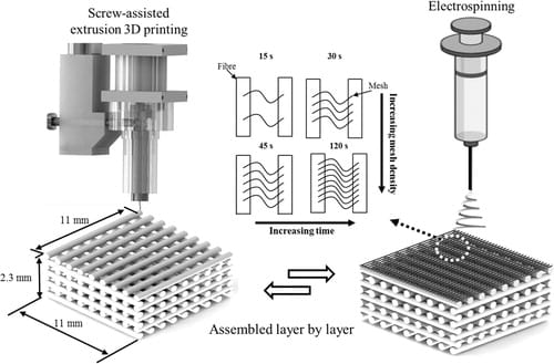
Using a 3D Discovery, screw-assisted extrusion 3D printer and an electrospinning system, the research team made dual-scale scaffolds with PCL, featuring a 0°/90° lay down, 300 μm pore size and fiber diameter, and 230 μm layer height. Processing parameters were as follows: .33 mm inner diameter (ID) nozzle, 90°C melt temperature, 12 mm/s deposition speed, and screw rate of 7.5 rpm. Fifteen sample scaffolds were printed.

Clinical Applications and Case Studies
Schematic representation of the 3D printing and electrospinning fabrication process of a dual-scale scaffold. 3D, three-dimensional.
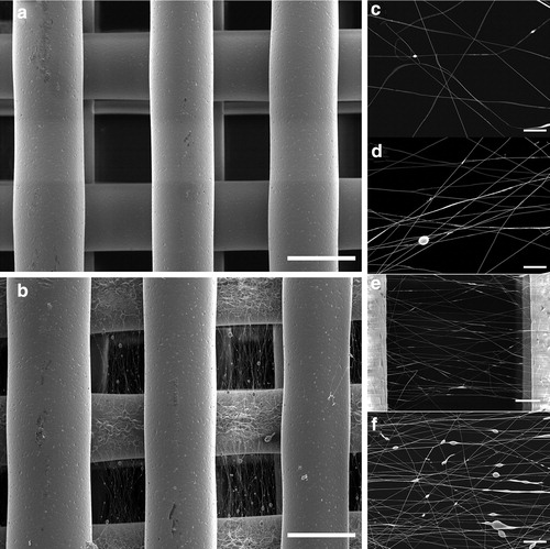
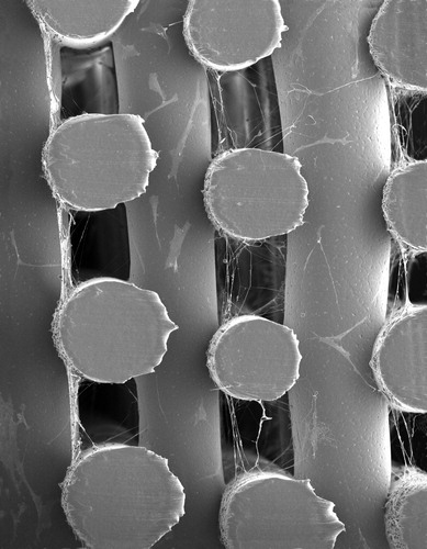
With a diameter of 820 ± 56 nm, electrospun fibers were integrated onto scaffolds, with beads displayed on the fibers and also in the pores. The researchers attributed the beads to possible issues with conductive properties, decrease in charge, or use of solvents. They did note that on the meshes fiber alignment was exhibited on those spun for 30 seconds or more.
Regulatory Considerations and Safety
SEM images of the (a) 3D-printed only scaffold and (b) dual-scale scaffold with electrospun (45 s) nanofibers (scale bar = 300 μm). Electrospun mesh density as function of time (c) 15 s, (d) 30 s, (e) 45 s, and (f) 120 s [scale bar = 20 μm, (e) 50 μm]. SEM, scanning electron microscopy.
Further analysis of the fibers confirmed a ‘clear preference’ for perpendicular alignment of electrospun fibers, connecting the printed fibers.
“The aligned fibers are present and homogeneously distributed throughout and within all the 3D-printed pores,” stated the researchers. “Furthermore, the electrospun fibers collected on the printed fibers themselves are oriented although not as clearly as the fibers within the pores.”
Research Breakthroughs and Innovations
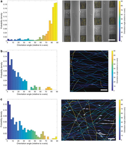
Fiber orientation analysis of SEM images. (a) Low magnification image showing aligned electrospun fibers (45 s) within all the pores of the printed scaffold (scale bar = 300 μm). Probability density of the orientation angle (relative to x-axis) shows a distinct distribution for angles between 75° and 90°. Higher magnification images (b) 45 s (scale bar = 50 μm) and (c) 120 s (scale bar = 20 μm) show aligned fibers with the orientation angle distributed toward 0°. Color images are available online.
“Further investigation is required to understand how the conductivity of the material influences fiber formation and alignment potentially through the incorporation of conductive fillers such as graphene or the use of conductive polymers. The electrical charge distribution can also be altered by changing the printed scaffold geometry (e.g., hexagonal and triangular) and incorporating both conductive and insulating regions within the structure to influence fiber alignment,” concluded the researchers. “This study is a promising development in the fabrication of multiscale scaffolds that better reflect the complexity of native tissue and the ability to engineer specific architectures to control cell behavior.”
What do you think of this news? Let us know your thoughts! Join the discussion of this and other 3D printing topics at 3DPrintBoard.com.
The Future of Bioprinting and Medical AM
[Source / Images: ‘Three-Dimensional Printing and Electrospinning Dual-Scale Polycaprolactone Scaffolds with Low-Density and Oriented Fibers to Promote Cell Alignment’]
The post University of Manchester: Improved Cell Alignment with 3D Printed & Electrospun PCL Scaffolds appeared first on 3DPrint.com | The Voice of 3D Printing / Additive Manufacturing.
Frequently Asked Questions
How is 3D printing used in medicine?
3D printing is used in medicine for surgical planning models, custom implants, bioprinting tissue scaffolds, drug delivery systems, dental aligners, and prosthetics. It enables patient-specific solutions that improve outcomes and reduce surgery time.
What materials are biocompatible for 3D printing?
Common biocompatible materials include PEEK, titanium alloys (Ti6Al4V), bio-ceramics (hydroxyapatite), medical-grade resins, PLA for temporary implants, and hydrogels for bioprinting. Material choice depends on the application and required mechanical properties.
Is 3D printed medical equipment FDA approved?
Yes, several 3D printed medical devices have FDA clearance, including orthopedic implants, dental restorations, and surgical guides. Each device must go through the appropriate regulatory pathway based on its risk classification.
📌 Related Articles
- Best Budget 3D Printer Upgrades That Actually Improve Print Quality: Belts, Springs, Hotends & More
- Best 3D Printer Upgrades That Actually Improve Print Quality: Complete 2026 Guide
- Prusa Research Mini+ vs Prusa MK4: Full Specs Comparison & Buyer’s Guide
- ABS 3D Printing Settings Guide: Temperature, Enclosure, and Cooling for Strong Parts
- Bambu Lab P1S vs Bambu Lab P2S: Full Specs Comparison & Buyer’s Guide