
Last month, doctors in east China’s Nanchang city used 3D printing technology for the first time to assist a complex cardiovascular surgery. Before outlining their final plan, a pre-operative evaluation team at the Second Affiliated Hospital of Nanchang University, in China’s Jiangxi Province, relied on 3D printing to simulate a transcatheter aortic valve replacement (TAVR) surgery on a high-risk elderly patient with severe aortic valve stenosis. Using 3D printing to conduct in vitro experimental simulations they were able to assess the risks related to the surgery. The successful operation led them to acknowledge that 3D printing technology could help other physicians design more personalized treatment solutions for their patients.
Aortic valve stenosis is a common clinical heart disease with high morbidity and mortality occurring when the aortic valve of the heart fails to open completely, preventing the normal flow of blood. Once symptoms appear, if surgical treatment is not performed, the mortality rate is very high, with one-year and five-year survival rates at 52% and 22%, respectively. It is the most common valvular heart disease of the elderly and increases with age. In China, the prevalence of moderate or severe aortic stenosis in patients more than 75 years old is over two percent.
As reported by the Second Affiliated Hospital, to treat the condition, surgeons usually perform a thoracotomy aortic valve replacement, but for the elderly and people with other serious underlying diseases, the surgical risk is much greater than the risk of the disease itself. Instead, surgeons tend to choose TAVR valve replacement surgery because it is minimally invasive, does not require extracorporeal circulation, and has a high rate of post-surgery recovery. Nevertheless, many complications still plague this procedure, such as annulus rupture, acute coronary opening obstruction, and paravalvular leakage.
The Chinese surgeons asserted that during the TAVR operation, they cannot directly look at the entire aortic root, nor can they cut the heart to observe the internal anatomical structure. So, to prepare for the surgery, they usually solely rely on computerized tomography (CT) scans, magnetic resonance imaging (MRI), and cardiac ultrasounds that only provide them with a two-dimensional image of the patient and a very limited field of vision.

3D model reconstruction for preoperative TAVR surgery using a Venus A valve (Credit: Second Affiliated Hospital at Nanchang University)
Compared to traditional planning, 3D printing technology
Compared to traditional planning, 3D printing technology has proven extremely useful in presurgical preparations, by converting the patient’s two-dimensional image plane data into a 1:1 full- size model that surgeons can use to visualize and standardize a very complex procedure. There is growing evidence that these 3D-printed models improve patient safety and accuracy by decreasing operative times.
Under the leadership of Yanqing Wu, director of the Department of Cardiovascular Medicine at the Second Affiliated Hospital, a pre-operative evaluation team decided to first conduct in vitro experimental simulations to assess the patient’s risks during surgery before determining the operation strategy.
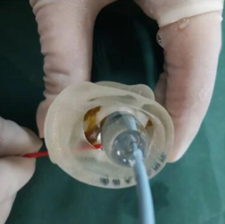
In vitro TAVR surgery simulation experiment (Credit: Second Affiliated Hospital at Nanchang University)
The 73-year-old male patient presented a real challenge for
The 73-year-old male patient presented a real challenge for surgeons. His physicians had detected a mid- to high-risk STS score, short for Society of Thoracic Surgery score, a validated risk-prediction model for open surgery based on data from the STS National Adult Cardiac Surgery Database. Additionally, preoperative CT scans from the patient suggested a bilobal aortic valve, severe calcification, and a high risk of coronary occlusion. Based on all these considerations, a 3D-printed presurgical approach was ideal.
Effectively, both the 3D reconstruction and the printed real images confirmed that the calcified lesions were worsening obstruction to the left ventricular outflow. The team then conducted an in vitro simulation experiment, using a 23-millimeter balloon for predilatation and a 26-millimeter TAVR Venus-A valve implant, from Venus Medtech (a China-based leader in medical devices).
The experimental results confirmed that after the valve was implanted, the left coronary artery opening was not affected in any way. Moreover, the results of the in vitro TAVR surgery simulation experiments proved predictable enough to move on to the patient surgery. Finally, with the cooperation of various professional teams, the patient’s TAVR operation was successfully completed.
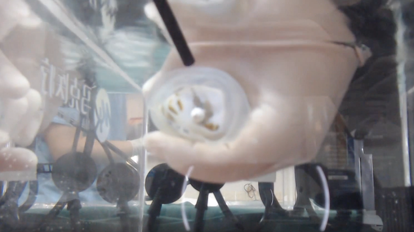
Surgeons simulate the TAVR surgery using a 3D printed model (Credit: Second Affiliated Hospital at Nanchang University)
The latest PARTNER 3 series of randomized controlled
The latest PARTNER 3 series of randomized controlled trials showed that in low-risk patients, TAVR was superior to another conventional surgical treatment of aortic valve stenosis known as surgical aortic valve replacement (SAVR) at reducing death, stroke, or rehospitalization at one year. Nonetheless, not all aortic stenosis patients are low-risk, in fact, the patient treated in Nanchang University’s hospital was deemed high-risk. Moreover, the experts at the hospital considered that patients, in general, should undergo a more thorough evaluation before surgery to truly achieve precision and individualized medicine.
This is a very different approach from the traditional treatment models based on unilateral know-how. In fact, this example of TAVR surgery based on presurgical 3D printing technology required effective teamwork in cardiovascular medicine, imaging experts, and cardiac surgeons to achieve successful results.
The hospital claims that the joint efforts of various professional disciplines and technology can help design highly personalized treatment plans for different patients, maximizing their safety. This is why the TAVR team of the Second Affiliated Hospital expects to lead future TAVR surgeries using 3D printing technology in the Jiangxi province to achieve more comprehensive and higher-quality progress.
The post 3D Printing for Preoperative Simulation of Complex Cardiovascular Surgery appeared first on 3DPrint.com | The Voice of 3D Printing / Additive Manufacturing.
from Your daily news from 3DPrint.com https://bit.ly/2VnA6K3